In-Office Testing
Lorem ipsum dolor sit amet, consectetur adipiscing elit. Nullam molestie velit arcu, a ullamcorper nibh pharetra in. Donec gravida eget nunc non vehicula. Nam id orci eu odio dignissim pulvinar in vitae orci. Praesent auctor odio diam, eget aliquet nisl sollicitudin at. Proin dignissim purus quam, sed dignissim ipsum malesuada eu. Nam pretium gravida lectus, vitae pharetra arcu ornare eu. Suspendisse sagittis finibus elit, a lacinia nunc rutrum sit amet. Mauris rhoncus id turpis a malesuada.
Lorem ipsum dolor sit amet, consectetur adipiscing elit. Nullam molestie velit arcu, a ullamcorper nibh pharetra in. Donec gravida eget nunc non vehicula. Nam id orci eu odio dignissim pulvinar in vitae orci. Praesent auctor odio diam, eget aliquet nisl sollicitudin at. Proin dignissim purus quam, sed dignissim ipsum malesuada eu. Nam pretium gravida lectus, vitae pharetra arcu ornare eu. Suspendisse sagittis finibus elit, a lacinia nunc rutrum sit amet. Mauris rhoncus id turpis a malesuada.
Lorem ipsum dolor sit amet, consectetur adipiscing elit. Nullam molestie velit arcu, a ullamcorper nibh pharetra in. Donec gravida eget nunc non vehicula. Nam id orci eu odio dignissim pulvinar in vitae orci. Praesent auctor odio diam, eget aliquet nisl sollicitudin at. Proin dignissim purus quam, sed dignissim ipsum malesuada eu. Nam pretium gravida lectus, vitae pharetra arcu ornare eu. Suspendisse sagittis finibus elit, a lacinia nunc rutrum sit amet. Mauris rhoncus id turpis a malesuada.
Lab Testing
Celiac disease (CD) is often undiagnosed and is caused in genetically predisposed individuals by abnormal intestinal permeability and abnormal immune response to gluten, a protein complex found in wheat, barley, spelt and rye. The inflammatory autoimmune response damages the lining of the small bowel and is associated with diarrhea, bloating, fatigue, nutritional deficiencies, and systemic autoimmune conditions. Gluten sensitivity can cause similar symptoms but without the same level of tissue damage. The Celiac & Gluten Sensitivity profile from Doctor’s Data helps differentiate between CD and gluten sensitivity by evaluating the serum titers of IgA and IgG for deamidated gliadin peptide, gliadin, and gluten.
This test is useful for:
- Patients who have persistent skin conditions (rash) or ataxia, idiopathic neurological conditions, autoimmune arthritis/ thyroiditis, unexplained weight loss or persistent gastrointestinal symptoms that are not associated with enteropathogens
- Symptomatic individuals that have tested positive for the HLA DQ2/DQ8 genotypes
- Patients with symptoms or symptom exacerbation with dietary gluten or re-introduction of gluten after a trial elimination of gluten
- Individuals that have a first degree relative with a diagnosis of CD
- Any child with a history of 3 or more antibiotic-treated cases of gastroenteritis while less than 6 months of age
- Patients on a gluten-inclusive diet who have Type I diabetes, Multiple Sclerosis or schizophrenia
- Individuals on a gluten-inclusive diet who have other laboratory evidence that may be associated with CD:
- Elevated liver function tests
- Bone demineralization
- Evidence of impaired absorption of fat-soluble vitamins, iron, B12 or folic acid
The Comprehensive Stool Analysis with Parasitology x1, 2, or 3 is an invaluable non-invasive diagnostic assessment that permits practitioners to objectively evaluate the status of beneficial and imbalanced commensal bacteria, pathogenic bacteria, yeast/fungus and parasites. Precise identification of pathogenic species and susceptibility testing greatly facilitates selection of the most appropriate pharmaceutical or natural treatment agents. Important information regarding the efficiency of digestion and absorption can be gleaned from the measurement of the fecal levels of elastase (pancreatic exocrine sufficiency), fat, muscle and vegetable fibers, and carbohydrates. Inflammation can significantly increase intestinal permeability and compromise assimilation of nutrients. The extent of inflammation, whether caused by pathogens or inflammatory bowel disease (IBD), can be assessed and monitored by examination of the levels of biomarkers such as lysozyme, lactoferrin, white blood cells and mucus. These markers can be used to differentiate between inflammation associated with potentially life-threatening inflammatory bowel disease (IBD), which requires lifelong treatment, and less severe inflammation that can be associated with irritable bowel syndrome (IBS) which is frequently due to the presence of enteroinvasive pathogens. Lactoferrin is only markedly elevated prior to and during the active phases of IBD, but not with IBS. Monitoring fecal lactoferrin levels in patients with IBD can therefore facilitate timely treatment of IBD, and the test can be ordered separately. Since the vast majority of secretory IgA (sIgA) is normally present in the GI tract, where it prevents binding of pathogens and antigens to the mucosal membrane, it is essential to know the status of sIgA in the gut. sIgA is the only bona fide marker of humoral immune status in the GI tract. Cornerstones of good health include proper digestion of food, assimilation of nutrients, exclusion of pathogens and timely elimination of waste. To obtain benefits from food that is consumed, nutrients must be appropriately digested and then efficiently absorbed into portal circulation. Microbes, larger-sized particles of fiber, and undigested foodstuffs should remain within the intestinal lumen. Poor digestion and malabsorption of vital nutrients can contribute to degenerative diseases, compromised immune status and nutritional deficiencies. Impairment of the highly specific nutrient uptake processes, or compromised GI barrier function, as in “leaky gut syndrome,” can result from a number of causes including:
- Low gastric acid production
- Chronic maldigestion
- Food allergen impact on bowel absorptive surfaces
- Bacterial overgrowth or imbalances (dysbiosis)
- Pathogenic bacteria, yeast or parasites and related toxic irritants
The use of NSAIDs and antibiotics Impairment of intestinal functions can contribute to the development of food allergies, systemic illnesses, autoimmune disease, and toxic overload from substances that are usually kept in the confines of the bowel for elimination. Efficient remediation of GI dysfunctions incorporates a comprehensive guided approach that should include consideration of elimination of pathogens and exposure to irritants, supplementation of hydrochloric acid, pancreatic enzymes and pre- and probiotics, and repair of the mucosal barrier.
This test measures the ability of two sugar molecules, lactulose and mannitol, to permeate the intestinal epithelial barrier. Ordinarily, mannitol is efficiently absorbed but lactulose, a larger molecule, is not. This test can help to identify malabsorption and “leaky gut” syndrome (abnormal intestinal permeability), which is often associated with inflammation specifically in the gastrointestinal tract. This test requires a baseline urine collection followed by a six-hour timed urine collection after ingesting a lactulose and mannitol solution.
This test is useful for
- Gastrointestinal Symptoms
- Food Sensitivities
- Inflammation
- Malabsorption
- Nutritional Deficiencies
Measurement of amino acids and diagnostic metabolites.
Red blood cell (RBC) analysis is an invaluable method for assessing insufficiency or excess of elements that have important functions within cells or on blood cell membranes. An important feature of this analysis is that the cells are not washed, because this would result in partial loss of some important elements, such as calcium, that bind to the plasma membrane.
RBC element levels are very useful for:
- Cardiotonic influences (magnesium, potassium)
- Anti-inflammatory processes (selenium, copper, zinc)
- Anemia (copper, iron)
- Immunological function (zinc, copper, magnesium)
- Glucose tolerance (chromium, manganese and possibly vanadium)
Disorders specifically associated with zinc deficiency are also addressed by this analysis. These disorders include loss of visual acuity, dysgeusia, dermatitis and poor wound healing, alopecia, amino acid malabsorption, sexual impotence, decreased production of testosterone, depressed immune function and growth retardation.
Accurate assessment of essential element status is highly recommended for the determination of appropriate supplementation. The absorption, transport and metabolism of essential elements is highly integrated and regulated. Inappropriate supplementation or dietary imbalance of elements can have significant adverse health effects. For example, excess intake of zinc or molybdenum can result in copper deficiency and, although essential, excess retention of manganese can have serious neurotoxic effects.
RBC element analysis is also useful for the assessment of ongoing or very recent exposure to specific toxic elements that accumulate preferentially in erythrocytes. These toxic elements include arsenic, cadmium, lead, methylmercury and thallium. It is important to keep in mind that elevated levels of the toxic elements in these cells reflect only recent or ongoing exposure and do not provide information about the net retention of the metals in the body.
RBC element analysis should be performed prior to and intermittently throughout the course of detoxification or chelation therapy. Monitoring essential element status is necessary to identify needs for and effectiveness of supplementation. Replacement and maintenance of adequate levels of essential nutrients can markedly reduce the apparent adverse “side effects” associated with the use of detoxification agents and the general effects of mobilization of toxic elements. It is important to note that some diseases are associated with abnormal levels of blood cell elements that could be misleading with respect to nutritional status. For example, blood cell copper can be temporarily elevated during inflammatory response while liver levels are not.
Many individuals have “hidden” impairments in amino acid metabolism that are problematic and often go undiagnosed. These impairments may or may not be expressed as specific symptoms. They may silently increase susceptibility to a degenerative disease or they may be associated with, but not causative for, a disease. Because of the wealth of information provided, it is suggested that a complete amino acid analysis be performed whenever a thorough nutritional and metabolic workup is called for.
Amino acid analysis provides fundamental information about nutrient adequacy, including the quality and quantity of dietary protein, digestive disorders, and vitamin and mineral deficiencies—particularly folic acid, B12, B6 metabolism, zinc and magnesium. In addition, amino acid analysis provides important diagnostic information about hepatic and renal function, availability of precursors of neurotransmitters, detoxification capacity, susceptibility to occlusive arterial disease (homocystine), and many inherent disorders in amino acid metabolism.
The patient’s results are presented in a functional format that permits ease of interpretation. A comprehensive summary of “presumptive needs” (such as B6, B12/folate, Mg) and “implied conditions” (such as maldigestion/malabsorption, abnormal gastrointestinal flora, impaired detoxification, oxidative stress) are presented based upon each patient’s results. Patient-specific amino acid supplement schedules and user-friendly commentary are provided to simplify nutritional intervention.
Plasma is traditionally used to assess the status of essential AA while urine analysis provides more information regarding AA wasting and aberrant metabolism associated with co-factor insufficiencies.
Plasma amino acid analysis measures what is being transported at the time of sampling. The specimen should be collected after an overnight fast to reduce the influence of dietary protein. Abnormalities are deduced by comparison of measured levels with an established reference range.
The 24-hour urine amino acid analysis has the highest probability of detecting abnormalities if renal function is normal. The 24-hour test indicates what is high and low over the course of a day, reflects blood and tissue amino acid pools, and is not affected by circadian rhythm. Healthy kidneys efficiently conserve essential amino acids. Therefore, urine levels of amino acids decrease first and tend to give an earlier indication of inadequacy than do plasma levels.
A first morning void urine (FMV) amino acid analysis, with results normalized per gram creatinine, provides an alternative when a complete 24-hour collection is not a viable option. The FMV analysis is excellent for identification of marked abnormalities, particularly with respect to gastrointestinal health, inherited disorders in amino acid metabolism and renal function, and can be used for protein challenge testing.
Urinary porphyrins are oxidized intermediate metabolites of heme biosynthesis and are readily excreted in excess when porphyrinogens accumulate as a result of inhibition of specific enzymes in the heme biosynthetic pathway. Heme is required for oxygen binding, transport and utilization, cytochromes, and electron transport in mitrochondira. The high rate of production of heme facilitates the use of urinary porphyrins as early and sensitive biomarkers of disorders in heme production, which has long been associated with genetic disorders, metabolic disturbances and diseases, nutritional status, oxidative stress and high-level exposure to toxic chemicals or metals.
Specific urinary porphyrin profiles have been associated with very high levels of toxic metals such as mercury (Hg), lead and arsenic. Mercury specifically inhibits two enzymes in porphyrinogen metabolism—uroporphyrinogen decarboxylase and coproporphyrinogen oxidase (CPOX). Inhibition of those two enzymes, particularly in the renal cortex, results in accumulation of pentacarboxyporphyrinogen and coproporphyrinogen III. Oxidation of the abnormally elevated porphyrinogens results in elevated urinary levels of total porphyrins, pentacarboxyporphyrin and coproporphyrin III. Recent research has identified an additional abnormal porphyrin in the urine of Hg-exposed dentists and also in rats fed very high levels of mercury for extended periods of time. A third Hg-associated porphyrin is most commonly referred to as “precoproporphyrin” as it elutes after pentacarboxyporphyrin and before coproporphyrin I. Precoproporphyrin has yet to be characterized with respect to molecular identity and appears to be elevated in Hg-exposed individuals who carry a variant of the CPOX gene (CPOX4 polymorphism).
Research at Doctor’s Data, Inc. has identified three separate precoproporphyrin peaks. Since knowledge about the Hg-associated precoproporphyrin entities is limited, we report the levels of all three peaks separately, as well as the total, for research use. Since uroporphyrin levels are not known to be affected by Hg, we also report the total precoproporphyrins-to-uroporphyrin ratio to increase the sensitivity for detecting abnormalities in individuals with low heme biosynthesis, as may occur in children with nutritional deficiencies or autism.
In addition:
- Arsenic exposure has been associated with elevated levels of uroporphyrins and coproporphyrin I, and an elevated ratio of coproporphyrins (I: III).
- Lead exposure has been associated with elevated levels of coproporphyrin III.
- Exposure to hexachlorobenzene and dioxin has been associated with elevated levels of uroporphyrins.
- Exposure to polyvinylchloride (PVC) and polybrominated biphenyl has been associated with elevated levels of coproporphyrins.
Various drugs and other substances can suppress enzymes involved in porphyrin metabolism and affect the levels of urinary porphyrins. Such compounds include alcohol, sedatives, analgesics, antibiotics, estrogens and oral contraceptives. Anemia, pregnancy and liver disease can also affect porphyrin metabolism.
This non-invasive test requires a single first morning void (FMV) or 24-hour urine collection.
Analysis of the levels of toxic metals in urine after the administration of a metal detoxification agent is an objective way to evaluate the accumulation of toxic metals. Acute metal poisoning is rare. More common, however, is a chronic, low-level exposure to toxic metals that can result in significant retention in the body that can be associated with a vast array of adverse health effects and chronic disease.
One cannot draw valid conclusions about adverse health effects of metals without assessing net retention. For an individual, toxicity occurs when net retention exceeds physiological tolerance. Net retention is determined by the difference between the rates of assimilation and excretion of metals. To evaluate net retention, one compares the levels of metals in urine before and after the administration of a pharmaceutical metal detoxification agent such as EDTA, DMSA or DMPS. Different compounds have different affinities for specific metals, but all function by sequestering “hidden” metals from deep tissue stores and mobilizing the metals to the kidneys for excretion in the urine.
It is important to perform both pre- and post-provocation urinalysis to permit distinction between ongoing exposures to metals (pre-) and net bodily retention. The pre-provocation urine collection can also be utilized to assess the rate of creatinine clearance if a serum specimen is also submitted.
Many clinicians also request the analysis of essential elements in urine specimens to evaluate nutritional status and the efficacy of mineral supplementation during metal detoxification therapy. Metal detoxification agents can significantly increase the excretion of specific nutrient elements such as zinc, copper, manganese and molybdenum.
Chromium metabolism authorities suggest that 24-hour chromium excretion likely provides the best assessment of chromium status. Early indication of renal dysfunction can be gleaned from urinary wasting of essential elements such as magnesium, calcium, potassium and sodium in an unprovoked specimen.
Variability in urine volume can drastically affect the concentration of elements. To compensate for urine dilution variation, elements are expressed per unit creatinine for timed collections. For 24-hour collections, elements are reported as both units per 24 hours and units per creatinine.
This test is useful for:
- Toxic Element Exposure
- Alopecia
- Bone Density
- Cardiovascular Disease
- Depression
- Dermatitis or Poor Wound Healing
- Detoxification Therapy
- Fatigue
- Gastrointestinal Symptoms
- Hypertension
- Immune Function
- Impaired Glucose Tolerance
- Inflammation
- Kidney Function
- Nutritional Deficiencies
- Parkinson’s-like Symptoms
Tests for minerals and metals found in the urine.
The thyroid evaluation panel tests all of the thyroid hormones, such as TSH, free T4, free T3, and reverse T3. It also tests for the antibodies working against the thyroid gland found in autoimmune diseases.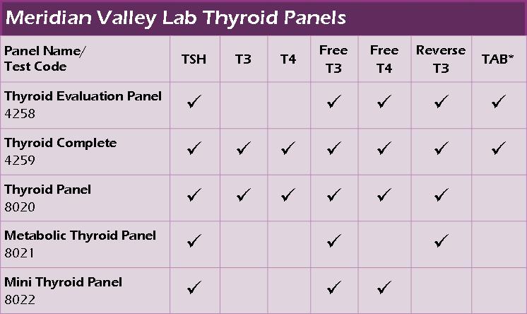
MRT is the only sensitivity blood test in the entire world that quantifies the degree of the inflammatory response in sensitivity pathways. That means in addition to identifying the foods with the highest degree of reaction, more importantly, MRT is able to identify the foods that have the lowest degree of reaction (in sensitivity pathways).
Immunologically speaking, the least reactive MRT foods are your patient’s best foods (as long as you have taken account of any food allergies and celiac disease). The least reactive MRT tested foods have the highest probability of being well-tolerated. And if they are foods the patient likes to eat, which they usually are, those are the foods that will form the basis for the multi-phased LEAP eating plan.
This feature of MRT eliminates the guesswork and makes building a healthy diet much easier. This is one of the main reasons we have such excellent practitioner and patient loyalty.
LEAP then builds a diet in a systematic and controlled way (phased approach), allowing the patient to implement their dietary changes with a great deal of success and satisfaction.
In an IgG reaction, the IgG antibodies attach themselves to the food antigen and create an antibody-antigen complex. These complexes are normally removed by special cells called macrophages. However, if they are present in large numbers and the reactive food is still being consumed, the macrophages can’t remove them quickly enough. The food antigen-antibody complexes accumulate and are deposited in body tissues. Once in tissue, these complexes release inflammation causing chemicals, which may play a role in numerous diseases and conditions.
Why Test IgG Food Sensitivity?
There is a growing body of evidence to support the clinical benefits of eliminating IgG reactive foods from the diet. IgG food sensitivities have been implicated in migraine headaches and irritable bowel syndrome (alternating diarrhea and constipation). Bloating and indigestion are also common food sensitivity reactions, as is fatigue. Continued consumption of reactive foods may contribute to weight gain and/or difficulty losing weight. Eczema is also commonly associated with food reactions. Because IgG food reactions take hours or days to develop, this makes it difficult to determine which food is responsible for the reaction without doing testing.
Toxic chemicals are found to varying degrees in everything from cosmetic and skin care products to cookware and children’s toys. Chronic exposure to the following pollutants may cause them to accumulate in the body and perhaps contribute to disease.
Phthalates are used in the manufacture of plastics. Soft plastics are known for releasing phthalates into the contents of the plastic container.
Parabens are chemical preservatives that prevent growth of bacteria and mold and increase the shelf life of many perishable products. Approximately 75% of skin products contain parabens.
Also known as volatile solvents, VOCs are air borne particles that contribute to poor air quality and smog.
Symptoms related to environmental pollutants may include:
- irritation of eyes, nose and throat; skin rashes and other skin irritations
- effects on the central nervous system including impaired thinking and movement
- disrupting the function of hormones
- headaches, nausea, anxiety, or depression
- fatigue and drowsiness
Why Get an Environmental Pollutants Profile?
Because the human body breaks down pollutants and eliminates them in the urine, measuring the metabolites of these pollutants in urine is an effective way to assess the level of exposure.
The Fatty Acid Profile measures the percentage of fatty acids in red blood cells from a convenient dried blood spot. Measurements include the Omega-3 Index and Omega-3 score to assess for heart disease risk, the ratio of Arachidonic Acid (AA) to Eicosapentaenoic acid (EPA) as a marker of inflammation, total Omega-3 fatty acids, total Omega-6 fatty acids along with mono-unsaturated fatty acids, trans fatty acids and saturated fats. Measurement of fatty acid content in red blood cell membranes shows less biological variability than measurement in plasma or serum.
The essential fatty acid family includes omega-3 and omega-6 fatty acids, which are considered essential because they only come from food. Most of us get plenty of omega-6 fatty acids from grains and grain fed animals, but our diets often lack omega-3 fatty acids. A few of the more important omega-3 fatty acids are: eicosapentaenoic acid (EPA), docosohexaenoic acid (DHA) and alpha linolenic acid (ALA). The right balance of omega-3 fatty acids has been proven to reduce the risk of heart attack, and having the right balance of omega-3 and 6 fatty acids may help reduce inflammation.
Trans fatty acids (TFAs or trans fats) are a chemically altered form of good fatty acids. Food manufacturers began altering fatty acids to increase stability to improve shelf life. Saturated fats occur naturally, usually in animal meat, and are solid or semi-solid at room temperature. Unfortunately, we now know that both trans fats and saturated fats raise the ‘bad’ LDL cholesterol and thus increase risk for heart disease.
Why Get a Fatty Acid Profile Test?
Preventing a heart attack starts with knowing which risk factors you have and correcting what you can. The Fatty Acid Profile helps by measuring your fatty acid levels and letting you know how your numbers stack up compared to a normal population. And, if you start supplementing with fish oils and avoiding those trans and saturated fats, you can always retest and see how well you’re doing.
Good health depends in part on having adequate amounts of essential nutritional elements, and low levels of toxic elements. Hair element analysis shows nutrient status (essential elements) and toxic element exposure for the length of time it took to grow the hair supplied for testing (usually about three months).
Good health depends in part on having adequate amounts of essential nutritional elements, and low levels of toxic elements. Hair element analysis shows nutrient status (essential elements) and toxic element exposure for the length of time it took to grow the hair supplied for testing (usually about three months).
Essential Elements
Essential elements are essential because they are needed to maintain health. For example, potassium and sodium are essential for proper heart function, while calcium and magnesium are essential for bone development,
Toxic Elements
Toxic elements usually cause damage by taking the place of essential elements and interfering with their usual function. For example, calcium helps strengthen bone, but lead can take the place of calcium in bone, making bone more porous and less sturdy.
Why Get Hair Element Analysis?
Hair element analysis is an easy and inexpensive way to assess the levels of essential and toxic elements. Less than one gram of hair is needed to test for 45 essential and toxic elements. Rocky Mountain Analytical provides a custom interpretation that explains the patterns of element levels in hair.
Lorem ipsum dolor sit amet, consectetur adipiscing elit, sed do eiusmod tempor incididunt ut labore et dolore magna aliqua. Ut enim ad minim veniam, quis nostrud exercitation ullamco laboris nisi ut aliquip ex ea commodo consequat. Duis aute irure dolor in reprehenderit in voluptate velit esse cillum dolore eu fugiat nulla pariatur. Excepteur sint occaecat cupidatat non proident, sunt in culpa qui officia deserunt mollit anim id est laborum.
Lorem ipsum dolor sit amet, consectetur adipiscing elit, sed do eiusmod tempor incididunt ut labore et dolore magna aliqua. Ut enim ad minim veniam, quis nostrud exercitation ullamco laboris nisi ut aliquip ex ea commodo consequat. Duis aute irure dolor in reprehenderit in voluptate velit esse cillum dolore eu fugiat nulla pariatur. Excepteur sint occaecat cupidatat non proident, sunt in culpa qui officia deserunt mollit anim id est laborum.
Lorem ipsum dolor sit amet, consectetur adipiscing elit, sed do eiusmod tempor incididunt ut labore et dolore magna aliqua. Ut enim ad minim veniam, quis nostrud exercitation ullamco laboris nisi ut aliquip ex ea commodo consequat. Duis aute irure dolor in reprehenderit in voluptate velit esse cillum dolore eu fugiat nulla pariatur. Excepteur sint occaecat cupidatat non proident, sunt in culpa qui officia deserunt mollit anim id est laborum.
A true food allergy is triggered by IgE antibody production specific to a reactive food. IgE reactions generally occur within minutes of eating a reactive food, which is why they are also called ‘immediate’ hypersensitivity reactions. After the first exposure to a food allergen, the body remembers what the allergen looks like and keeps a supply of IgE ready for immediate release if it sees that allergen again. Food allergies can be life-threatening (for example, an anaphylactic reaction to peanuts), but these reactions are rare, occurring in less than 1% of people. Skin reactions like hives and eczema, plus breathing and digestive problems are also common IgE reactions. Referral to an allergy specialist is recommended in the case of serious food allergies.
The IgE panel offered by Rocky Mountain Analytical may be useful for identifying IgE reactions to regularly eaten foods responsible for unexplained symptoms like:
- abdominal pain
- diarrhea
- eczema
- heartburn
- nausea
- vomiting
Why Test for IgE Food Allergy?
Serious reactions like difficulty breathing or anaphylactic reactions should be diagnosed and treated by a physician or a healthcare professional trained in treatment of allergic reactions. However, otherwise unexplained and chronic symptoms like those listed above may be signs of food allergies.
Lorem ipsum dolor sit amet, consectetur adipiscing elit, sed do eiusmod tempor incididunt ut labore et dolore magna aliqua. Ut enim ad minim veniam, quis nostrud exercitation ullamco laboris nisi ut aliquip ex ea commodo consequat. Duis aute irure dolor in reprehenderit in voluptate velit esse cillum dolore eu fugiat nulla pariatur. Excepteur sint occaecat cupidatat non proident, sunt in culpa qui officia deserunt mollit anim id est laborum.
Lorem ipsum dolor sit amet, consectetur adipiscing elit, sed do eiusmod tempor incididunt ut labore et dolore magna aliqua. Ut enim ad minim veniam, quis nostrud exercitation ullamco laboris nisi ut aliquip ex ea commodo consequat. Duis aute irure dolor in reprehenderit in voluptate velit esse cillum dolore eu fugiat nulla pariatur. Excepteur sint occaecat cupidatat non proident, sunt in culpa qui officia deserunt mollit anim id est laborum.
Lorem ipsum dolor sit amet, consectetur adipiscing elit, sed do eiusmod tempor incididunt ut labore et dolore magna aliqua. Ut enim ad minim veniam, quis nostrud exercitation ullamco laboris nisi ut aliquip ex ea commodo consequat. Duis aute irure dolor in reprehenderit in voluptate velit esse cillum dolore eu fugiat nulla pariatur. Excepteur sint occaecat cupidatat non proident, sunt in culpa qui officia deserunt mollit anim id est laborum.
Lorem ipsum dolor sit amet, consectetur adipiscing elit, sed do eiusmod tempor incididunt ut labore et dolore magna aliqua. Ut enim ad minim veniam, quis nostrud exercitation ullamco laboris nisi ut aliquip ex ea commodo consequat. Duis aute irure dolor in reprehenderit in voluptate velit esse cillum dolore eu fugiat nulla pariatur. Excepteur sint occaecat cupidatat non proident, sunt in culpa qui officia deserunt mollit anim id est laborum.
Lorem ipsum dolor sit amet, consectetur adipiscing elit, sed do eiusmod tempor incididunt ut labore et dolore magna aliqua. Ut enim ad minim veniam, quis nostrud exercitation ullamco laboris nisi ut aliquip ex ea commodo consequat. Duis aute irure dolor in reprehenderit in voluptate velit esse cillum dolore eu fugiat nulla pariatur. Excepteur sint occaecat cupidatat non proident, sunt in culpa qui officia deserunt mollit anim id est laborum.
Elevated IgG antibodies to Candida are often found when someone has a fungal infection of some sort. For example, vaginal yeast infection, ‘jock itch’, and ringworm. Consuming too many sugar-rich foods can contribute to overgrowth of Candida. Symptoms associated with Candida overgrowth include:
- clouded thinking
- depression
- diarrhea
- fatigue
- menstrual pain
- vaginal yeast infections
- headaches
Chronic exposure to Candida, particularly when it has colonized or invaded the gut lining, can result in the elevation of IgG, IgA, and IgM specific antibodies.
The Candida Panel measures levels of IgG, IgA and IgM antibodies to Candida plus Candida antigen in a blood strip sample. Measurement of Candida antigen in blood indicates widespread infection, which can occur in immunocompromised individuals.
Lorem ipsum dolor sit amet, consectetur adipiscing elit, sed do eiusmod tempor incididunt ut labore et dolore magna aliqua. Ut enim ad minim veniam, quis nostrud exercitation ullamco laboris nisi ut aliquip ex ea commodo consequat. Duis aute irure dolor in reprehenderit in voluptate velit esse cillum dolore eu fugiat nulla pariatur. Excepteur sint occaecat cupidatat non proident, sunt in culpa qui officia deserunt mollit anim id est laborum.
Cortisol is the major Adrenal corticosteroid, also referred to as hydrocortisone. Some Important functions include regulation of blood pressure, immune function, cardiovascular function, as well as regulation of metabolism relating to proteins, carbohydrates, and fats. Cortisol is strongly bound to Corticosteroid Binding Globulin in the circulation, with smaller amounts loosely bound to Albumin and some unbound (free form). Cortisol production is under negative feedback from the pituitary/hypothalamus via Adrenocorticotropic hormone and Corticotropin Releasing Factor. It exhibits diurnal variation with highest values early in the morning (6-8 am) and the nadir at 11pm. This diurnal variation is absent in Cushing’s syndrome and also disrupted by number of factors. Cortisol is also called the “stress” hormone because levels increase in response to stressful factors, whether physiological/physical (illness, trauma, surgery etc) or psychological. Measurements of Cortisol levels help diagnose Cushing syndrome and Addison disease, the two major adrenal disorders. However, they are also sometimes used for evaluating possible psychological or stress-related conditions.
DHEA-S is a steroid derived almost exclusively (90%) from the adrenal gland. It is the sulfated form of DHEA, and its concentration in serum is greater than that of any other steroid hormone and is about five hundred times that of DHEA. DHEA-S is not subject to diurnal variations nor changes during the menstrual cycle. This is partly due to its slow metabolic clearance as a result of binding to albumin. DHEA-S levels are highest at age 20-25 and decline with advancing years, reaching 20-30% of peak levels by age 70-80. It is a weak androgen and commonly used for assessment of adrenal androgen production. Concentrations of DHEAS are often measured, along with other hormones such as FSH, LH, prolactin, estrogen, and testosterone, to help diagnose polycystic ovarian syndrome (PCOS) and to help rule out other causes of infertility, amenorrhea, and hirsutism. This test is used also used as an aid in the evaluation of androgen excess, adrenocortical disease including congenital adrenal hyperplasia, and adrenal tumor. More recently, DHEA-S has been suggested to be a possible mediator in response to stress and perhaps a new marker of low libido in women under 45 years (rather than testosterone).
Estradiol is the most biologically active form of all the naturally occurring estrogens and the most important one. In females, during reproductive years, it is largely is produced by the ovaries. Smaller amounts are also produced by the adrenal glands and from metabolism of testosterone in the peripheral circulation. Estradiol is responsible for development of sex organs and characteristics in females. It is also found in males, but in much smaller concentrations.
Estradiol circulates mostly bound to estrogen binding globulin (also referred to as SHBG) with a smaller fraction loosely bound to albumin. A small percentage circulates unbound to proteins (free form). The free and weakly bound are considered biologically active forms. Estradiol levels are most commonly used to evaluate ovarian function, and in males to diagnose the cause of gynecomastia. Estradiol is also used to monitor menopausal hormone replacement therapy.
Progesterone is a steroid hormone derived from pregnenolone. In females, it is produced mostly by the ovaries. The production is higher during the luteal phase (second half) of the menstrual cycle and during pregnancy. It is low during the follicular phase (first half), before puberty, and after menopause. The primary biological function is to prepare the uterus to provide nutrition to the developing embryo during early pregnancy. Progesterone is also used for menopausal hormone replacement therapy and for women with infertility or frequent pregnancy loss. Measurement of progesterone is generally performed to evaluate infertility, corpus luteum function, basal body temperature (for monitoring ovulation), abnormal bleeding, placental function in pregnancy, and monitor some women on hormone replacement therapy.
Testosterone is the major androgen responsible for sexual differentiation and male secondary sex characteristics. In the circulation, approximately 55% is tightly bound to sex hormone binding globulin (SHBG), 42% to albumin, and about 3% is free (unbound). The free and about half of the albumin fraction is bioactive. In males, testosterone is primarily secreted by the testes and levels exhibit diurnal variation with the highest levels between 6 and 9 AM and lowest around midnight. Testosterone levels begin to decline with age starting at about 40 yrs. In females, about half of the testosterone is derived from peripheral conversion of androstenedione. Major applications of testosterone measurement reside with assessment of sexual development, gonadal disorders, hyperandrogenic syndromes, menstrual disorders, and hormone replacement therapy.
The Comprehensive Stool Analysis detects the presence of pathogenic microorganisms such as yeast, parasites, and bacteria that contribute to chronic illness and neurological dysfunction. It provides helpful information about prescription and natural products effective against specific strains detected in the sample. The test also evaluates beneficial bacteria levels, intestinal immune function, overall intestinal health, and inflammation markers.
Many chronic disorders come from digestive problems and inadequate nutrient absorption. Proper gastrointestinal function is needed to eliminate toxic substances, pathogenic microbes, and undigested food particles from the body to prevent health problems. Nutrients require a specific internal environment to be properly digested and transported throughout the body.
Abnormal intestinal microorganisms in the GI tract are widely known to cause disease. Research shows a relationship between the GI tract and the neurological, hepatic, and immune systems. For example, excessive yeast produces toxic substances that can pass through the blood-brain barrier and alter neurological functioning causing “brain fog,” behavior problems, and learning difficulties.
The Organic Acids Test (OAT) provides an accurate evaluation of intestinal yeast and bacteria. Abnormally high levels of these microorganisms can cause or worsen behavior disorders, hyperactivity, movement disorders, fatigue and immune function. Many people with chronic illnesses and neurological disorders often excrete several abnormal organic acids. The cause of these high levels could include: oral antibiotic use, high sugar diets, immune deficiencies, and genetic factors.
If abnormalities are detected using the OAT, treatments can include supplements, such as vitamins and antioxidants, or dietary modification. Upon treatment, patients and practitioners have reported significant improvement such as decreased fatigue, regular bowel function, increased energy and alertness, increased concentration, improved verbal skills, less hyperactivity, and decreased abdominal pain. The OAT is strongly recommended as the initial screening test.
The Microbial Organic Acids Test (MOAT) is ideal for follow-up to the OAT and is often recommended by practitioners looking for a specific abnormality, to monitor certain microbial imbalances, or to assess treatment efficacy.
This panel provides a comprehensive view of adrenal function and includes 4 cortisol levels timed throughout the day as well as DHEA. Symptoms that would indicate ordering this panel include:
- Fatigue
- Nervousness
- Weakness
- Sugar cravings
- Insomnia
- Dizzy spells
- Headaches
- Decreased stamina
- Irritability
- Fibromyalgia
- Anxiety/Depression
Saliva testing is an easy and noninvasive way of assessing your patients hormone balancing needs, and is proving to be the most reliable medium for measuring hormone levels. Unlike serum tests, saliva testing represents only hormones actively delivered to receptors in the body. Clinically, it is far more relevant to test these bioavailable hormones and provide an accurate reflection of the body’s active hormone levels.
The Comprehensive Hormone Panel is the starting point for initial assessment of hormonal status and endocrine function. This panel is useful with male and female patients because it looks at the full diurnal cortisol pattern. This panel tests the following hormones: Estradiol (E2), Progesterone, Testosterone, DHEA, a.m. Cortisol, noon Cortisol, evening Cortisol, p.m. Cortisol.
The charge is $45 per hormone tested/$360 for the 8 hormones. Results are emailed in a PDF document. One of College Pharmacy’s pharmacists will help walk you through the process once your Saliva Test Kit order has been placed.
*Note: All saliva samples are frozen and kept for 25 days beyond receipt to enable additional testing. If preliminary values indicate a more comprehensive panel, missing hormones can be added.
Supplement Information
- Labrix™ saliva testing is an easy non-invasive way of assessing your hormone balance. This information allows you to supplement hormones according to a plan tailored for only you. Well-balanced hormones regulate everything from reproduction to emotions, general health and well-being. An imbalance of any one hormone can throw your physical and mental health out of balance, causing aggravating and even serious health problems.
- Labrix uses one of the most advanced processes available for accurately assessing hormone levels in saliva. Labrix uses four saliva samples, timed throughout the day, for results that accurately reflect your hormonal status, as some hormones levels vary somewhat during the course of the day. Most other testing labs use just one saliva sample collected in the morning.
- Why do all four tubes need to be submitted if only one test is ordered? For the testing of Estradiol, DHEA, Progesterone, and Testosterone, we take an equal amount of specimen from each tube and transfer it into a 5th tube, the pool, which is used for testing. This process ensures a more accurate overall average of the hormone level, compensating for the natural daily peaks and troughs of these analytes. If only one tube is collected at a random time of day, there is no way to know if the result reflects one of those daily fluctuations and is accurate.
Neurotransmitter imbalances can be easily identified with a single noninvasive urine sample. Testing provides a tool to understand each patient’s specific neuroendocrine imbalances, which can be corrected with targeted nutritional therapy, BHRT, diet, and lifestyle interventions. Because it is especially important to understand the interrelationships of the neurotransmitters as well as their relationships with adrenal and sex hormones, an optimal approach measures each of the six neurotransmitter levels identified here in addition to a full hormone panel.
NeuroHormone Complete Panel Plus Neurotransmitter Test, Hormone, and Adrenal Test
The Neuroendocrine Connection: Adrenal hormones, sex hormones, and neurotransmitters are functionally interrelated. Changes in sex hormones and adrenal hormones can lead to neurotransmitter imbalances. In turn, neurotransmitter imbalances can affect hormone function. Labrix offers neurotransmitter/hormone combination panels that can provide a more comprehensive view of the body’s functional neuroendocrine status, and can bring to light additional factors contributing to symptoms. This panel provides a more comprehensive look into the status of the sex hormones, as well as neurotransmitters and adrenal function.
The NeuroHormone Complete Plus panel includes estrone (E1) and estriol (E3) plus the estrogen quotient. The addition of neurotransmitters to the Comprehensive Plus hormone only panel provides insight on how HPA axis function may be contributing to symptom manifestation such as mood swings, fatigue, and pain. Because the research on the estrogen quotient and the protective properties of estriol has not been done with men, this panel is currently recommended for women only.